

Pancreatic Neuroendocrine Tumor (PanNET) Successfully Treated with Laparoscopic Distal Pancreatectomy
Overview
Pancreatic neuroendocrine tumors (PanNETs) are uncommon tumors that arise from the hormone-producing (endocrine) cells of the pancreas.
Unlike the more common pancreatic adenocarcinoma, many PanNETs grow slowly and may produce hormones that lead to distinctive clinical symptoms.
Early recognition and timely treatment can significantly improve outcomes.
This clinical case describes the successful management of a 33-year-old gentleman who presented with recurrent episodes of severe hypoglycaemia (low blood sugar) resulting in transient loss of consciousness.
Over several months, he experienced repeated unexplained attacks despite having no known history of diabetes.
These symptoms prompted detailed evaluation, which revealed a small, well-defined tumor in the tail of the pancreas.
Contrast-enhanced CT scan and endoscopic ultrasound (EUS) identified a localized pancreatic lesion, and EUS-guided biopsy confirmed a well-differentiated pancreatic neuroendocrine tumor.
After multidisciplinary evaluation, the patient underwent laparoscopic distal pancreatectomy with splenectomy with the goal of complete tumor removal.
Histopathological examination demonstrated a well-differentiated Grade 2 pancreatic neuroendocrine tumor (pT2) with tumor-free surgical margins and no lymphovascular or perineural invasion.
Immunohistochemistry supported the diagnosis, and subsequent Ga-68 DOTATATE PET-CT surveillance showed no evidence of residual disease, recurrence, or distant metastasis during long-term follow-up.
The patient’s hypoglycaemic episodes resolved completely after surgery, and he returned to normal daily activities.
This case illustrates the importance of recognizing unexplained hypoglycaemia as a potential manifestation of a functional pancreatic neuroendocrine tumor.
It also demonstrates how accurate diagnosis, multidisciplinary planning, minimally invasive surgery, detailed pathological assessment, and structured long-term surveillance can contribute to excellent clinical outcomes.
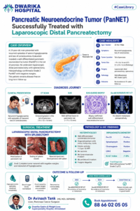
Case Highlights
| Parameter | Details |
| Patient Age | 33 years |
| Gender | Male |
| Presenting Symptoms | Recurrent hypoglycaemia with episodes of transient loss of consciousness |
| Duration of Symptoms | Approximately 3 months |
| Primary Diagnosis | Well-differentiated Pancreatic Neuroendocrine Tumor (PanNET) |
| Probable Functional Status | Functional pancreatic neuroendocrine tumor presenting with hypoglycaemia |
| Tumor Location | Tail of pancreas |
| Tumor Size on CT | Approximately 19 × 10 × 16 mm |
| Tumor Size on Histopathology | Approximately 2.1 × 2.0 × 1.6 cm |
| Preoperative Tissue Diagnosis | EUS-guided biopsy confirmed neuroendocrine tumor |
| Surgical Procedure | Laparoscopic distal pancreatectomy with splenectomy |
| Histological Grade | Grade 2 well-differentiated neuroendocrine tumor |
| Pathological Stage | pT2 |
| Surgical Margins | Negative (R0 resection) |
| Lymphovascular Invasion | Not identified |
| Perineural Invasion | Not identified |
| Immunohistochemistry | Positive for Chromogranin, Synaptophysin, and Pan CK; Ki-67 approximately 7–8% |
| Postoperative Outcome | Resolution of hypoglycaemic episodes and uneventful recovery |
| Long-Term Follow-up | Serial Ga-68 DOTATATE PET-CT scans showed no recurrence or metastasis |
| Current Clinical Status | Disease-free on follow-up with good functional recovery |
Patient Presentation
How did the patient first present?
A 33-year-old gentleman presented with a history of recurrent episodes of sudden unconsciousness over the preceding three months. These episodes were found to be associated with marked hypoglycaemia, raising concern for an underlying endocrine disorder rather than a primary neurological condition.
Along with these episodes, the patient reported a weight gain of approximately 8 kilograms over three months. He did not complain of abdominal pain, jaundice, fever, anorexia, or other gastrointestinal symptoms. There was no documented history of diabetes mellitus, and he had no history of smoking or alcohol consumption.
The recurrent nature of the hypoglycaemic attacks significantly affected his daily life and prompted referral for specialist evaluation. Because unexplained hypoglycaemia in a non-diabetic adult is uncommon, a detailed clinical assessment was undertaken to determine the underlying cause.
Key Presenting Features
- Recurrent episodes of hypoglycaemia
- Transient loss of consciousness
- Approximately three months’ duration of symptoms
- Weight gain of around 8 kg
- No jaundice
- No abdominal pain
- No fever
- No anorexia
- No significant history of tobacco or alcohol use
Why were these symptoms important?
Persistent or recurrent hypoglycaemia in an adult without diabetes warrants careful evaluation. While several medical conditions can cause low blood sugar, one important possibility is a functional pancreatic neuroendocrine tumor, particularly an insulin-secreting tumor (insulinoma). Such tumors release excessive insulin, resulting in repeated episodes of low blood glucose that may present with sweating, palpitations, confusion, behavioural changes, seizures, or loss of consciousness.
Clinical Evaluation
How was the diagnosis suspected?
The patient’s presentation of recurrent hypoglycaemia in the absence of diabetes immediately raised suspicion for an endocrine cause of hypoglycaemia.
Given the repeated episodes of transient unconsciousness and the absence of more common explanations, further evaluation focused on identifying a possible insulin-producing pancreatic lesion.
The initial clinical assessment aimed to determine:
- Whether the hypoglycaemia had an endocrine origin.
- Whether there was evidence of a pancreatic lesion.
- Whether the lesion appeared localized or had spread beyond the pancreas.
- Whether surgical treatment would be feasible if a tumor was identified.
To answer these questions, the patient underwent a structured diagnostic work-up that included high-quality cross-sectional imaging followed by endoscopic ultrasound and tissue diagnosis.
Differential diagnoses considered
Based on the patient’s presentation, the differential diagnosis of recurrent hypoglycaemia could include:
- Functional pancreatic neuroendocrine tumor
- Drug-induced hypoglycaemia
- Endocrine disorders affecting glucose regulation
- Critical systemic illness
- Rare metabolic causes
Clinical reasoning
Several features increased the likelihood of a localized pancreatic neuroendocrine tumor:
- Recurrent symptomatic hypoglycaemia in a young adult.
- Absence of diabetes mellitus.
- No obvious alternative cause identified
- Subsequent imaging demonstrating a discrete lesion in the tail of the pancreas.
These findings supported further investigation with dedicated pancreatic imaging and tissue diagnosis.
Investigations & Imaging Findings
Contrast-Enhanced CT (CECT) Abdomen
A contrast-enhanced CT scan of the abdomen demonstrated a well-defined enhancing lesion in the tail of the pancreas, measuring approximately 19 × 10 × 16 mm. The lesion showed imaging characteristics suggestive of a pancreatic neuroendocrine tumor.
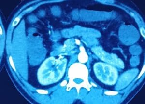
Importantly, the CT scan also demonstrated:
- No significant dilatation of the pancreatic duct.
- No evidence of major vascular invasion.
- No significant abdominal lymphadenopathy.
- The remaining pancreas appeared unremarkable.
These findings suggested a localized pancreatic lesion without obvious radiological evidence of advanced disease, supporting consideration of curative surgical resection.
Endoscopic Ultrasound (EUS)
To obtain more detailed assessment of the pancreatic lesion, the patient underwent endoscopic ultrasound (EUS).
EUS identified a well-defined lesion in the tail of the pancreas measuring approximately 17 × 13 mm. Because EUS provides high-resolution imaging of the pancreas and allows tissue sampling, an EUS-guided biopsy was performed during the same procedure.
EUS-Guided Biopsy
Histological examination of the biopsy specimen demonstrated features consistent with a well-differentiated neuroendocrine tumor, providing tissue confirmation before surgery. This diagnosis, combined with the clinical presentation and imaging findings, enabled informed multidisciplinary discussion and surgical planning.
Final Diagnosis
Following clinical evaluation, imaging, and tissue diagnosis, the patient was diagnosed with a localized well-differentiated pancreatic neuroendocrine tumor involving the tail of the pancreas.
The diagnosis was supported by:
- Characteristic clinical presentation with recurrent hypoglycaemia.
- Contrast-enhanced CT showing a localized enhancing pancreatic tail lesion.
- Endoscopic ultrasound confirming the lesion.
- EUS-guided biopsy demonstrating a well-differentiated neuroendocrine tumor.
Why Was Surgery Recommended?
Why was surgery considered the best treatment option?
Following detailed clinical evaluation, imaging, and tissue diagnosis, the multidisciplinary team concluded that surgical removal offered the best opportunity for definitive treatment in this patient.
The lesion was confined to the tail of the pancreas, showed no radiological evidence of distant spread, and biopsy confirmed a well-differentiated pancreatic neuroendocrine tumor (PanNET).
The patient’s repeated episodes of severe hypoglycaemia significantly affected his quality of life and carried the risk of serious complications such as accidents, seizures, or permanent neurological injury if left untreated.
Because these symptoms were attributable to a localized pancreatic neuroendocrine tumor, complete surgical excision offered the greatest likelihood of resolving the underlying cause.
For localized, resectable pancreatic neuroendocrine tumors, surgery is generally regarded as the preferred treatment with curative intent.
In this case, the combination of clinical presentation, imaging findings, and biopsy confirmation supported proceeding with minimally invasive surgical resection.
What factors supported surgical treatment?
Several findings favoured surgery:
- Young patient with good performance status.
- Localized tumor in the tail of the pancreas.
- No evidence of distant metastasis on preoperative evaluation.
- No major vascular involvement on CT imaging.
- Histological confirmation of a well-differentiated neuroendocrine tumor by EUS-guided biopsy.
- Symptomatic disease with recurrent hypoglycaemia.
- Feasibility of complete tumor removal using a minimally invasive approach.
Expected goals of surgery
The objectives of surgery were to:
- Completely remove the pancreatic tumor.
- Eliminate the source of recurrent hypoglycaemia.
- Preserve long-term quality of life.
- Achieve tumor-free surgical margins (R0 resection).
- Reduce the risk of future disease progression or recurrence.
As demonstrated during follow-up, these objectives were successfully achieved in this patient.
Surgical Planning
How was the operation planned?
Successful pancreatic surgery begins long before entering the operating room. Careful planning was undertaken to determine whether the tumor could be safely removed while minimizing surgical risks.
Imaging-based planning
The contrast-enhanced CT scan provided several important pieces of information:
- Exact location of the lesion within the pancreatic tail.
- Small tumor size (approximately 19 × 10 × 16 mm).
- No pancreatic duct dilatation.
- No obvious vascular invasion.
- No significant regional lymphadenopathy.
These findings suggested that complete surgical removal through a minimally invasive approach was technically feasible.
Tissue diagnosis before surgery
Unlike many pancreatic tumors that proceed directly to surgery based on imaging alone, this patient underwent EUS-guided biopsy, which confirmed a well-differentiated neuroendocrine tumor before the operation. This helped establish the diagnosis and supported surgical decision-making.
Choice of surgical approach
The surgical team planned a laparoscopic distal pancreatectomy with splenectomy.
The planned operation aimed to:
- Remove the entire tumor with adequate margins.
- Remove the distal pancreas containing the lesion.
- Achieve complete oncological clearance.
- Perform the procedure using minimally invasive techniques whenever safely feasible.
Surgical Challenges
What challenges were anticipated before surgery?
Although the tumor was relatively small, pancreatic surgery requires meticulous planning because of the pancreas’ complex anatomy and close relationship with major blood vessels and adjacent organs.
Based on the location and available records, the anticipated technical challenges included:
1. Deep retroperitoneal location
The pancreatic tail lies deep within the upper abdomen, making exposure technically demanding during minimally invasive surgery.
2. Relationship to splenic vessels
The distal pancreas is closely associated with the splenic artery and splenic vein. Safe dissection requires careful identification and control of these vessels.
3. Achieving complete tumor removal
The surgical objective was complete excision with negative margins while preserving surrounding structures whenever appropriate.
4. Prevention of postoperative pancreatic fistula
After distal pancreatectomy, secure closure of the pancreatic remnant is essential to reduce the risk of leakage of pancreatic enzymes.
5. Management of a functional endocrine tumor
Because the patient’s symptoms were related to recurrent hypoglycaemia, complete tumor removal was essential to eliminate the source of abnormal hormone production and resolve the metabolic disturbance.
Operative Strategy
What operation was performed?
The patient underwent:
Laparoscopic Distal Pancreatectomy with Splenectomy.
Why was a laparoscopic approach chosen?
A minimally invasive laparoscopic approach offers several potential advantages in appropriately selected patients, including:
- Smaller incisions.
- Reduced postoperative pain.
- Earlier mobilisation.
- Faster recovery.
- Shorter hospital stay.
- Improved cosmetic outcome.
Principles of the operation
The operative strategy included:
- Access to the upper abdomen using a minimally invasive approach.
- Identification of the distal pancreas.
- Careful mobilisation of the pancreatic tail.
- Safe management of the splenic vessels.
- Complete removal of the distal pancreas together with the spleen.
- Retrieval of the specimen for pathological examination.
These steps are consistent with the documented surgical procedure, although individual technical details are not available in the uploaded records.
What was found during surgery?
Tumor located in the distal pancreas close to the posterior surface.
- Well-circumscribed lesion in distal pancreas. No adhesions, vascular anatomy normal. No operative difficulty, 50 ml blood loss, operative duration 2 hrs and no intraoperative complications.
What did the pathology report show?
Histopathological examination confirmed successful complete removal of the tumor.
The resected specimen consisted of distal pancreatectomy with splenectomy. Gross examination identified a well-circumscribed solid tumor in the distal pancreas measuring approximately 2.1 × 2.0 × 1.6 cm.
Microscopic examination
Microscopy demonstrated:
- Well-differentiated neuroendocrine tumor.
- Organoid architecture with nests, trabeculae, cords, ribbons, and glandular patterns.
- Uniform tumor cells with finely stippled (“salt-and-pepper”) chromatin.
- Rich vascular network.
- Mitotic activity of 1–2 per 10 high-power fields.
- No tumor necrosis.
Surgical margins
All examined resection margins were free of tumor, indicating complete excision (R0 resection).
Lymphovascular and perineural invasion
The pathology report demonstrated:
- No lymphovascular invasion.
- No perineural invasion.
Pathological stage
According to the pathology report:
- Well-differentiated Pancreatic Neuroendocrine Tumor
- Grade 2
- Pathological stage: pT2
Immunohistochemistry (IHC)
Immunohistochemical staining further confirmed the diagnosis:
| Marker | Result |
| Pan CK | Positive |
| Synaptophysin | Positive |
| Chromogranin | Positive |
| CDX2 | Negative |
| Ki-67 Index | Approximately 7–8% |
These findings supported the diagnosis of a well-differentiated Grade 2 pancreatic neuroendocrine tumor.
Why are these pathology findings important?
Several pathological features indicate a favourable outcome in this case:
- Complete tumor removal with negative margins.
- Well-differentiated tumor biology.
- Absence of lymphovascular invasion.
- Absence of perineural invasion.
- No tumor necrosis.
- Localized disease (pT2).
- Immunohistochemistry confirming neuroendocrine differentiation.
These findings, together with the patient’s excellent postoperative course and disease-free follow-up, support the success of surgical treatment in this case.
Postoperative Care
How was the patient cared for after surgery?
Successful pancreatic surgery does not end in the operating room. Careful postoperative monitoring is essential to identify complications early, support recovery, and ensure safe discharge.
Blood Sugar Monitoring
Because the patient initially presented with recurrent symptomatic hypoglycaemia, careful postoperative glucose monitoring was particularly important.
During follow-up:
- Fasting blood glucose remained within the normal range.
- Postprandial blood glucose levels remained satisfactory.
- Most importantly, the patient experienced complete resolution of recurrent hypoglycaemic attacks after surgery.
This clinical improvement strongly suggested successful removal of the hormone-producing tumour responsible for the patient’s symptoms.
Nutritional Recovery
Follow-up consultations documented encouraging nutritional recovery.
The patient reported:
- Good appetite
- Progressive improvement in general health
- Stable body weight during later follow-up
- No significant dietary intolerance related to surgery.
Following distal pancreatectomy, gradual progression from liquids to a normal balanced diet is typically recommended according to individual recovery and medical advice.
Recovery During Follow-up Visits
- No recurrence of hypoglycaemic episodes
- Normal random blood sugar
- Good appetite
- Stable weight
- Occasional constipation managed conservatively
- No significant abdominal complaints
- Overall satisfactory postoperative recovery.
Outcome & Recovery
What was the outcome after surgery?
The patient’s postoperative outcome was excellent.
Following laparoscopic distal pancreatectomy with splenectomy:
- Hypoglycaemic episodes resolved completely.
- Loss of consciousness did not recur.
- Daily blood sugar values remained stable.
- Appetite improved.
- Functional recovery was satisfactory.
- The patient resumed routine activities.
This represents the primary objective of surgery in a patient presenting with a functional pancreatic neuroendocrine tumour causing recurrent hypoglycaemia.
Improvement in Quality of Life
Before surgery, recurrent hypoglycaemic episodes significantly disrupted the patient’s daily life and posed potential risks such as accidents and neurological injury.
Following tumour removal, the patient’s quality of life improved considerably.
Clinic records over subsequent years consistently documented:
- Feeling well
- No recurrent hypoglycaemia
- No significant abdominal pain
- No bloating
- No nausea or vomiting
- Stable health during routine follow-up.
Were there any documented postoperative complications?
There was no major postoperative complications such as:
- Pancreatic fistula
- Intra-abdominal abscess
- Reoperation
- Postoperative haemorrhage
- Delayed gastric emptying
Long-Term Follow-up
Why is long-term follow-up important after pancreatic neuroendocrine tumour surgery?
Even after successful surgery, patients with pancreatic neuroendocrine tumours require structured follow-up because recurrence can occasionally occur months or years after treatment.
Long-term surveillance helps:
- Detect recurrence early.
- Identify distant metastasis if present.
- Assess long-term metabolic recovery.
- Monitor nutritional status.
- Evaluate quality of life.
Clinical Follow-up
The patient continued regular follow-up over 4 years till the writing of this article.
During these visits, the records consistently documented:
- No recurrence of hypoglycaemic episodes.
- Stable body weight.
- Good appetite.
- Overall well-being.
- No significant abdominal complaints.
- No evidence of clinical disease recurrence.
Ga-68 DOTATATE PET-CT Surveillance
One of the strengths of this case is the availability of serial functional imaging.
Follow-up PET-CT (2023)
Ga-68 DOTATATE PET-CT demonstrated:
- No somatostatin receptor (SSTR)-expressing lesion at the operative site.
- No loco-regional lymph node metastasis.
- No distant metastatic disease.
- No evidence of recurrent tumour
Follow-up PET-CT (2024)
Repeat Ga-68 DOTATATE PET-CT again demonstrated:
- No recurrent disease.
- No lymph node metastasis.
- No distant metastasis.
- Stable postoperative findings.
Most Recent Clinical Follow-up
The most recent outpatient review documented:
- Patient feeling well.
- Stable weight.
- No abdominal pain.
- No nausea or vomiting.
- No constipation requiring significant intervention.
- Continued disease-free status.
Long-Term Outcome
This case demonstrates several favourable long-term outcomes:
- Complete symptom resolution.
- Excellent postoperative recovery.
- Disease-free follow-up over more than three years.
- No evidence of local recurrence.
- No evidence of distant metastasis.
- Excellent functional outcome.
- Return to normal life.
Timeline of Care
| Timeline | Clinical Event |
| 3 months before diagnosis | Recurrent hypoglycaemic episodes with transient loss of consciousness |
| Initial evaluation | Clinical assessment raised suspicion of an endocrine pancreatic lesion |
| Contrast-enhanced CT | Enhancing tumour identified in the tail of the pancreas (approximately 19 × 10 × 16 mm) |
| Endoscopic Ultrasound (EUS) | Well-defined pancreatic tail lesion confirmed |
| EUS-guided biopsy | Well-differentiated pancreatic neuroendocrine tumour diagnosed |
| Multidisciplinary evaluation | Surgical treatment planned |
| 23 June 2022 | Laparoscopic distal pancreatectomy with splenectomy performed |
| Histopathology | Grade 2 well-differentiated pancreatic neuroendocrine tumour (pT2), negative margins |
| Immunohistochemistry | Pan CK, Synaptophysin and Chromogranin positive; Ki-67 approximately 7–8% |
| 2023 Follow-up | Ga-68 DOTATATE PET-CT showed no recurrence or metastasis |
| 2024 Follow-up | Repeat PET-CT remained disease free |
| Latest Clinical Review | Patient clinically well with no recurrence of hypoglycaemia and good functional recovery |
Surgical Pearls
This case highlights several practical lessons in pancreatic surgery:
- Recurrent unexplained hypoglycaemia in a non-diabetic adult should prompt evaluation for a functional pancreatic neuroendocrine tumour.
- High-quality pancreatic imaging is essential for accurate localisation of small pancreatic lesions.
- Endoscopic ultrasound with tissue diagnosis can assist preoperative planning in selected patients.
- Careful patient selection allows successful minimally invasive pancreatic surgery.
- Complete surgical excision with tumour-free margins remains the cornerstone of treatment for localized pancreatic neuroendocrine tumours.
- Histopathological grading and immunohistochemistry provide important prognostic information.
- Structured long-term surveillance, including functional imaging when appropriate, helps confirm durable disease control.
- Multidisciplinary management contributes to favourable oncological and functional outcomes.
Learning Points
- Functional pancreatic neuroendocrine tumours may present with recurrent hypoglycaemia rather than abdominal pain.
- Small pancreatic tumours can produce significant clinical symptoms.
- Cross-sectional imaging combined with endoscopic ultrasound improves diagnostic accuracy.
- Tissue diagnosis may assist treatment planning in selected patients.
- Minimally invasive distal pancreatectomy is an effective option for appropriately selected localized tumours.
- Complete tumour removal can resolve hormone-related symptoms.
- Histopathological grading influences prognosis and follow-up planning.
- Immunohistochemistry confirms neuroendocrine differentiation.
- Long-term surveillance is important even after apparently curative surgery.
- Early diagnosis and timely specialist referral can significantly improve patient outcomes.
Learning for Patients & Family
What can patients and families learn from this case?
Every patient with a pancreatic neuroendocrine tumour (PanNET) has a unique clinical presentation, and treatment decisions should always be individualised. This case highlights how early recognition of symptoms, appropriate investigations, specialist evaluation, and timely surgery can lead to excellent long-term outcomes.
1. Do not ignore recurrent episodes of low blood sugar
Repeated episodes of sweating, dizziness, confusion, fainting, seizures, or unconsciousness—especially in someone without diabetes—should never be considered normal. Prompt medical evaluation is essential because these symptoms may occasionally be caused by a hormone-producing pancreatic tumour.
2. Early diagnosis creates more treatment opportunities
Small pancreatic tumours are often easier to remove completely before they spread. Early diagnosis can increase the likelihood of curative treatment and reduce the need for more complex therapies.
3. Not every pancreatic tumour is pancreatic cancer
Many patients assume that every pancreatic tumour behaves aggressively. In reality, pancreatic neuroendocrine tumours differ from pancreatic adenocarcinoma in their biology, treatment options, and prognosis. Many localized PanNETs can be successfully treated with surgery.
4. Seek care from specialists experienced in pancreatic surgery
Treatment planning for pancreatic tumours often requires input from a multidisciplinary team that may include pancreatic surgeons, gastroenterologists, radiologists, pathologists, endocrinologists, oncologists, and anaesthesiologists. Experience in managing these uncommon tumours helps guide appropriate investigations and treatment.
5. Do not rely solely on internet or social media information
Online resources can be useful for learning, but they cannot replace an individual medical assessment. Treatment recommendations should always be based on:
- Your symptoms
- Clinical examination
- Blood investigations
- Imaging findings
- Biopsy (when appropriate)
- Overall health
- Discussion with your treating specialist
6. A second opinion is reasonable
If you have been advised to undergo major pancreatic surgery or have been diagnosed with a rare pancreatic tumour, obtaining a second opinion from an experienced pancreatic surgeon or multidisciplinary centre can help you better understand your diagnosis and treatment options.
7. Follow-up remains important even after successful surgery
Successful surgery does not eliminate the need for follow-up. Depending on the tumour type and pathology, your doctor may recommend:
- Clinical reviews
- Blood tests
- CT or MRI scans
- Functional imaging (such as Ga-68 DOTATATE PET-CT in selected patients)
Regular follow-up helps detect recurrence early and monitor long-term health. In this case, serial follow-up imaging showed no evidence of recurrence or metastasis during long-term surveillance.
8. Recovery extends beyond the operation
Recovery involves more than wound healing. Good nutrition, gradual return to physical activity, adherence to medical advice, and emotional support from family members all contribute to long-term well-being.
9. Stay hopeful
A diagnosis of a pancreatic tumour can be overwhelming. However, this case demonstrates that some pancreatic neuroendocrine tumours can be treated successfully when diagnosed early and managed appropriately. Every patient’s situation is different, but advances in imaging, minimally invasive surgery, pathology, and follow-up have improved outcomes for many people.
Learning for Referring Doctors / Primary Care Physicians
Key clinical messages for frontline healthcare providers
Early recognition and timely referral play a critical role in the management of pancreatic neuroendocrine tumours. This case provides several practical lessons for primary care physicians and referring clinicians.
1. Recognise red-flag symptoms
Consider endocrine causes when evaluating:
- Recurrent unexplained hypoglycaemia
- Episodes of altered consciousness
- Neuroglycopenic symptoms
- Recurrent symptoms relieved by glucose administration
Persistent symptoms warrant specialist evaluation.
2. Maintain a broad differential diagnosis
In patients with recurrent hypoglycaemia, consider:
- Functional pancreatic neuroendocrine tumour
- Drug-induced hypoglycaemia
- Endocrine disorders
- Metabolic causes
- Critical illness
The uploaded records confirm the diagnosis of a pancreatic neuroendocrine tumour but do not detail the complete differential diagnosis considered during evaluation.
3. Appropriate imaging is essential
High-quality pancreatic imaging helps determine:
- Tumour location
- Tumour size
- Relationship to adjacent structures
- Presence of vascular involvement
- Presence of metastatic disease
This information directly influences treatment planning.
4. Endoscopic ultrasound has an important role
For selected pancreatic lesions, EUS provides:
- High-resolution imaging
- Accurate localisation
- Tissue diagnosis through biopsy
In this case, EUS-guided biopsy confirmed a well-differentiated neuroendocrine tumour before surgery.
5. Early referral improves treatment options
Patients with suspected pancreatic tumours should be referred promptly to centres experienced in pancreatic disease. Delayed referral may postpone diagnosis and potentially reduce treatment options.
6. Multidisciplinary decision-making improves patient care
Management decisions benefit from collaboration among:
- Pancreatic surgeons
- Gastroenterologists
- Radiologists
- Pathologists
- Endocrinologists
- Medical oncologists
- Nuclear medicine specialists (when indicated)
7. Pathology determines prognosis
Important pathological parameters include:
- Tumour grade
- Ki-67 index
- Surgical margins
- Lymphovascular invasion
- Perineural invasion
- TNM stage
These findings help guide prognosis and postoperative surveillance.
8. Long-term surveillance should not be overlooked
Even after apparently curative surgery, structured follow-up remains important because recurrence may occur years later. The duration and intensity of surveillance should be individualised according to tumour biology, pathological findings, and current clinical guidelines.
9. Evidence-based takeaway
This case illustrates how careful clinical assessment, appropriate imaging, preoperative tissue diagnosis, minimally invasive surgery, detailed histopathological evaluation, and structured follow-up together contributed to an excellent long-term outcome.
Frequently Asked Questions (FAQ)
1. What is a pancreatic neuroendocrine tumour (PanNET)?
A pancreatic neuroendocrine tumour is an uncommon tumour that arises from the hormone-producing cells of the pancreas. Some PanNETs produce excess hormones and cause symptoms such as low blood sugar, while others are non-functional and may be detected incidentally.
2. Why did this patient experience recurrent hypoglycaemia?
According to the clinical records, the patient presented with recurrent hypoglycaemia and was subsequently diagnosed with a pancreatic neuroendocrine tumour. This clinical presentation is consistent with a functional pancreatic neuroendocrine tumour.
3. Why was laparoscopic surgery chosen?
The lesion was localised to the tail of the pancreas without documented distant spread, making laparoscopic distal pancreatectomy an appropriate surgical approach in this case. The minimally invasive technique can offer faster recovery and less postoperative discomfort in appropriately selected patients.
4. What did the pathology report show?
The tumour was a well-differentiated Grade 2 pancreatic neuroendocrine tumour (pT2) with negative surgical margins and no lymphovascular or perineural invasion. Immunohistochemistry supported the diagnosis.
5. Did the surgery cure the patient’s symptoms?
The uploaded records document complete resolution of the patient’s recurrent hypoglycaemic episodes after surgery, with continued clinical well-being during long-term follow-up.
6. Can this tumour come back?
Recurrence is possible in some patients with pancreatic neuroendocrine tumours, which is why regular follow-up is important. In this case, serial Ga-68 DOTATATE PET-CT scans demonstrated no evidence of recurrence or metastasis during the documented follow-up period.
7. What follow-up tests may be required after surgery?
Follow-up plans are individualised but may include:
- Clinical examination
- Blood tests
- CT or MRI scans
- Functional imaging (such as Ga-68 DOTATATE PET-CT when indicated)
The exact schedule should be determined by the treating specialist.
8. What factors influence prognosis in pancreatic neuroendocrine tumours?
Important factors include:
- Tumour size
- Histological grade
- Ki-67 index
- Tumour stage
- Completeness of surgical removal
- Presence or absence of metastasis
- Long-term follow-up findings
Related Disease Knowledge Hub
- Pancreatic Neuroendocrine Tumours (PanNETs)
- Functional Pancreatic Neuroendocrine Tumours
- Insulinoma
- Pancreatic Tumours
- Neuroendocrine Tumours of the Digestive System
Related Treatment & Service Pages
- Laparoscopic Distal Pancreatectomy
- Pancreatic Neuroendocrine Tumour Surgery
- Minimally Invasive Pancreatic Surgery
- Pancreatic Cancer Surgery
- Advanced Pancreatic Surgery
Educational Disclaimer
This clinical case is shared for educational purposes only. Patient-identifying information has been omitted to protect confidentiality. The clinical findings, investigations, treatment decisions, and outcomes described are specific to this individual case and should not be interpreted as the expected course for every patient.
Every patient is unique. The diagnosis and management of pancreatic neuroendocrine tumours depend on multiple factors, including symptoms, tumour biology, imaging findings, pathology, overall health, and multidisciplinary clinical assessment. Treatment recommendations should always be individualised after consultation with an experienced specialist.
The information presented in this Clinical Case Library is intended to improve understanding of pancreatic neuroendocrine tumours and their management. It is not a substitute for professional medical advice, diagnosis, or treatment. Patients experiencing symptoms or seeking guidance regarding a pancreatic condition should consult a qualified healthcare professional for personalised evaluation and care.
Conclusion
This case demonstrates how early recognition of recurrent hypoglycaemia, a structured diagnostic pathway using CT imaging, endoscopic ultrasound, and tissue diagnosis, followed by laparoscopic distal pancreatectomy with splenectomy, resulted in complete symptom resolution and sustained disease-free follow-up. The documented pathology, immunohistochemistry, and serial Ga-68 DOTATATE PET-CT surveillance make this an excellent example of multidisciplinary management of a localized pancreatic neuroendocrine tumour and provide valuable educational insights for patients, families, and healthcare professionals alike.

Super-specialist GI, bariatric & cancer surgeon. SGPGIMS (India's premier GI centre) + advanced training in Japan & South Korea. Read full profile →
Last reviewed: July 2026





Comments
Be the first to share your thoughts on this article.