


Omental Infarction: Causes & Treatment | A 17-Year-Old Girl Case Capsule. Recurrent Abdominal Pain, Managed Conservatively
#CaseCapsule | Omental Infarction
#ExpertRead | Acute Abdomen in Adolescents
Can omental infarction recur—or can residual infarct remain symptomatic?
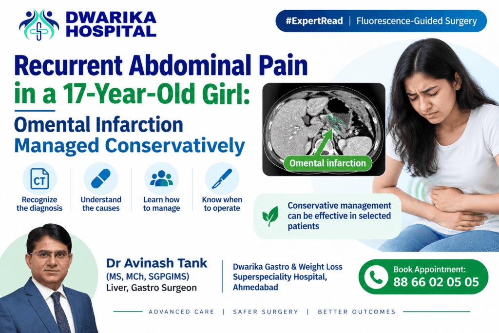
A 17-year-old girl presented with abdominal pain and inflammatory markers. Contrast-enhanced CT demonstrated a focal omental infarction. She improved with conservative treatment, and serial ultrasound showed reduction in the lesion. She subsequently developed recurrent abdominal pain, although her inflammatory markers were now normal.
This case highlights an important practical question:
When a young patient with radiologically diagnosed omental infarction develops recurrent pain, should we continue conservative treatment—or operate?
The Case
Patient: 17-year-old girl
Initial presentation
- Abdominal pain for approximately 20 days
- WBC: 14,000/µL
- CRP: 15
- CECT abdomen: focal omental infarction
- Admitted for observation and medical treatment
- Clinical improvement occurred without surgery
- Serial ultrasonography demonstrated progressive reduction of the omental lesion, reaching approximately 2 cm
The subsequent episode is clinically interesting.
Re-presentation
The patient developed abdominal pain again for 2 days.
At this time:
- WBC: normal
- CRP: normal
- Repeat CECT: focal omental lesion in the gastrocolic ligament, immediately below the gastric antrum, measuring approximately 23 × 13 mm
- No ascites
- No significant lymphadenopathy
- No bowel obstruction or significant bowel-wall abnormality
- Appendix normal
- No adnexal lesion
- Major abdominal vessels normal.
The radiologist’s impression was focal omental infarction.
An important imaging observation
The current CT should not automatically be interpreted as progression of the infarct.
The current lesion measures 23 × 13 mm, whereas the earlier CT described a larger, approximately 17 × 30 × 35 mm inflammatory omental lesion. Differences in CT plane, morphology and organization of an infarct can make direct size comparison difficult.
The important clinical finding is that there is no current leukocytosis or CRP elevation and no CT evidence of complication.
What exactly is omental infarction?
Omental infarction is ischemic necrosis of a segment of the greater omentum.
It occurs when the vascular supply to a portion of the omentum becomes compromised.
There are two broad mechanisms:
Primary omental infarction
Usually related to vascular compromise within the omentum, without an identifiable secondary disease.
Secondary omental infarction
May occur because of:
- Omental torsion
- Adhesions
- Hernia
- Previous abdominal surgery
- Trauma
- Local inflammatory disease
- Vascular abnormalities
- Hypercoagulable states
Torsion and thrombotic/vascular mechanisms are recognized pathways leading to omental ischemia.
Why is this diagnosis important in a young patient?
Omental infarction is uncommon, but it is increasingly recognized in children and adolescents.
It can mimic:
- Acute appendicitis
- Cholecystitis
- Gastritis/peptic disease
- Epiploic appendagitis
- Mesenteric adenitis
- Gastroenteritis
- Ovarian torsion
- Ruptured ovarian cyst
- Other causes of acute abdomen
In pediatric studies, omental infarction has frequently been associated with overweight/obesity, although it can certainly occur in patients without these risk factors.
What should we look for on CT?
Typical CT features include:
- Focal area of inflamed omental fat
- Heterogeneous fat attenuation
- Hyperattenuating streaks
- Peripheral hyperattenuating rim
- Surrounding inflammatory fat stranding
- Usually no bowel-wall pathology explaining the symptoms
The current CT in this case localizes the lesion to the gastrocolic ligament just below the antrum, measuring 23 × 13 mm.
The report also specifically documents:
- Normal appendix
- No significant bowel abnormality
- No free fluid
- No lymphadenopathy
- Normal major abdominal vessels.
These findings strengthen the diagnosis of an isolated omental process rather than appendicitis or another obvious intra-abdominal inflammatory disease.
The important question: Why did the pain recur?
This is where the case becomes educational.
Recurrent pain does not necessarily mean recurrent omental infarction.
There are several possibilities.
1. Persistent inflammatory change
Continue Reading — Free
Get the complete expert guide from Dr. Avinash Tank.
Free, always. No payment, no spam.
Continue Reading — Free
Get the complete expert guide from Dr. Avinash Tank.
Free, always. No payment, no spam — just the complete guide sent straight to your screen.
An infarcted segment of omentum can take time to completely resolve. The residual inflammatory mass may remain visible on ultrasound or CT even after the patient’s systemic inflammatory response has normalized.
2. Residual/reorganizing infarct
The reduction from the original lesion to approximately 2 cm on serial ultrasound supports an evolving/resolving process.
3. Omental torsion
A torsed omental segment can infarct and produce persistent or recurrent localized pain.
When reviewing the CT, specifically look for:
Whirling/convergence of omental vessels toward a vascular pedicle.
A definite whirl/pedicle sign makes torsion more clinically relevant.
4. A new episode
A genuinely new infarction is possible, although it should not be assumed simply because the patient has recurrent pain.
5. Another diagnosis
This is particularly important in a 17-year-old girl.
Recurrent abdominal pain should prompt reassessment for:
- Ovarian torsion
- Ovarian cyst
- Dysmenorrhea/endometriosis
- Gastritis/peptic disease
- Appendicitis
- Mesenteric pathology
- Urinary pathology
The current CT reported no adnexal lesion and a normal appendix, which is reassuring.
Management:
Conservative treatment remains the first option in an uncomplicated case
When CT establishes omental infarction and the patient is clinically stable, non-operative management is reasonable.
Treatment is principally supportive:
- Analgesia
- Hydration
- Antiemetics when required
- Oral diet as tolerated
- Serial abdominal examination
- Clinical observation
- Repeat CBC/CRP when clinically indicated
The pediatric literature supports conservative management when the diagnosis is secure and the child/adolescent is clinically stable. Several pediatric series have reported successful non-operative treatment without complications.
Ultrasound can also be useful for follow-up. In one pediatric series, serial ultrasound demonstrated disappearance of the infarction in all conservatively managed patients.
But when should we operate?
This is the most important surgical lesson.
Omental infarction itself is not an indication for surgery.
The decision should be based on the patient’s clinical course + imaging + diagnostic certainty.
Consider diagnostic laparoscopy when:
Persistent or worsening pain
Pain remains severe despite adequate conservative treatment, particularly if it is relentless.
Peritoneal signs
Guarding, rebound tenderness or progressive localized peritonism should lower the threshold for surgery.
Clinical deterioration
- Fever
- Rising WBC
- Rising CRP
- Tachycardia
- Sepsis
- Hemodynamic instability
Diagnostic uncertainty
If appendicitis, ovarian torsion, bowel pathology or another surgical condition cannot confidently be excluded.
Definite omental torsion
A clear twisted omental pedicle/whirl sign, particularly when associated with significant persistent pain, makes operative treatment more reasonable.
Pediatric surgical literature has specifically described uncertain diagnosis, intractable/relentless pain and persistent peritoneal findings as indications for operative exploration.
What does the evidence tell us about conservative versus surgical treatment?
There is still no universally accepted treatment guideline for omental infarction.
A systematic review of CT-diagnosed adult cases found that conservative treatment was used in approximately 73% of patients, with successful symptom resolution in approximately 84% of those treated conservatively.
However, the evidence has important limitations:
- Most studies are retrospective.
- Many are case reports or small series.
- Pediatric and adult populations are different.
- There is no high-quality randomized trial establishing one treatment as universally superior.
A pediatric series similarly supports a short trial of conservative management, while identifying persistent/intractable pain and peritoneal findings as reasons to operate.
A recent literature review also emphasizes intractable pain and omental torsion as situations in which surgery becomes more important.
What about this particular 17-year-old?
This case has several features favoring continued conservative management, provided the clinical examination is reassuring:
Favourable features
- Previous clinical improvement with medical treatment
- Serial USG demonstrated reduction of the lesion
- Current WBC is normal
- Current CRP is normal
- No free fluid
- No bowel obstruction
- No bowel-wall abnormality
- Normal appendix
- No adnexal lesion
- No abscess or collection
- Normal major abdominal vessels.
Therefore:
Recurrent pain alone, in the presence of normal inflammatory markers and an uncomplicated CT appearance, does not automatically justify omentectomy.
The next step should be clinical reassessment and careful review of the CT for evidence of omental torsion.
Should we investigate thrombophilia?
This is an important question in a 17-year-old.
Not routinely in every isolated omental infarction.
I would consider further evaluation when there are additional clues such as:
- Recurrent objectively documented infarction
- Thrombosis at another site
- Previous unexplained venous/arterial thrombosis
- Strong family history of thrombosis
- Unusual thrombotic events
- Clinical suspicion of a systemic hypercoagulable state
The mechanism of OI can involve hypercoagulable states or vascular abnormalities, but this does not mean that every young patient with OI has thrombophilia.
The Surgical Learning Point
Do not operate on the CT image. Operate on the patient.
A radiologically confirmed omental infarction in a stable adolescent with improving symptoms can usually be observed.
The trajectory is more important than the mere presence of an omental mass.
Think in three questions:
Is the diagnosis secure?
↓
Is the patient clinically improving or deteriorating?
↓
Is there evidence of torsion or another surgical pathology?
This approach prevents unnecessary surgery while maintaining an appropriate threshold for laparoscopy when the clinical course demands it.
Practical Decision Algorithm
17-year-old + localized abdominal pain
↓
US/CECT demonstrates omental infarction
↓
Stable + diagnosis secure + no peritonitis
→ Conservative treatment
→ Analgesia + hydration ± antiemetic
→ Clinical observation
→ Serial US/clinical follow-up when appropriate
↓
Improving
→ Continue conservative management
↓
Persistent severe pain / worsening examination
OR
Rising inflammatory markers
OR
Peritonitis
OR
Diagnostic uncertainty
OR
Definite omental torsion
→ Diagnostic laparoscopy ± resection of infarcted omentum
Key Learning Lessons from This Case
• Omental infarction should be included in the differential diagnosis of acute abdominal pain in adolescents.
• It can mimic appendicitis and other common surgical conditions.
• CT can establish the diagnosis and potentially prevent unnecessary surgery.
• Ultrasound is valuable not only for diagnosis but also for follow-up in children and adolescents.
• A decreasing lesion on serial ultrasound supports an evolving/resolving process.
• Normal WBC and CRP during recurrent pain are reassuring but do not replace clinical examination.
• Always review the CT specifically for an omental whirl/torsion sign.
• In a young female, ovarian pathology must remain in the differential diagnosis.
• Routine thrombophilia testing is not justified solely because the patient is young; recurrent or systemic thrombotic features should guide further evaluation.
• Persistent, relentless pain, peritonitis, diagnostic uncertainty or definite torsion are the main reasons to consider laparoscopy.
Bottom line
For a clinically stable 17-year-old with CT-proven uncomplicated omental infarction, conservative management with active clinical surveillance is a reasonable first-line strategy. Recurrent pain should trigger reassessment—not automatic surgery. The decision to operate should be driven by the clinical course, peritoneal findings, inflammatory response, diagnostic uncertainty and evidence of omental torsion.
Related patient videos
 DWARIKA HOSPITALVerified Patient Story
Verified
DWARIKA HOSPITALVerified Patient Story
Verified  DWARIKA HOSPITALVerified Patient Story
Verified
DWARIKA HOSPITALVerified Patient Story
Verified  DWARIKA HOSPITALVerified Patient Story
Verified
DWARIKA HOSPITALVerified Patient Story
Verified 
Related treatments & services
A complete, personalised weight-loss programme — surgery where needed, plus nutrition, medical and lifestyle support for lasting results.
View service → 360 Diabetes ReversalA complete programme to reverse Type-2 diabetes — combining metabolic surgery where suitable with diet, medical and lifestyle care.
View service → Gallbladder Stone
Minimally invasive removal of the gallbladder (laparoscopic cholecystectomy) — the definitive, day-care treatment for painful gallstones.
View service →Was this guide helpful?
Super-specialist GI, bariatric & cancer surgeon. SGPGIMS (India's premier GI centre) + advanced training in Japan & South Korea. Read full profile →
Last reviewed: July 2026